- X-ray is still the most common veterinary imaging tool in 2025. It handles 60-65% of cases. But ultrasound, CT, and MRI are each best for specific problems.
- Cost varies a lot: X-rays cost $150-$400. MRI scans cost $2,000-$3,500. Choosing the right test the first time often saves money. You won’t need to repeat imaging.
- Advanced imaging isn’t always necessary. Understanding when to move from basic X-rays to CT or MRI helps. It prevents missing problems and unnecessary costs.
Your vet might talk about “Veterinary Diagnostic Imaging 2025: X-Ray vs Ultrasound vs CT vs MRI Compared.” It can sound confusing.
But here’s the thing. I’ve watched pet owners struggle with these choices. They wonder if that $2,000 MRI is really necessary. Maybe a $300 X-ray would work instead.
Or worse, they choose the cheaper test. Then they need the expensive imaging anyway. This happens after weeks of uncertainty.
The veterinary imaging world has changed a lot. Understanding which tool does what matters. It affects your pet’s outcome and your wallet.
The veterinary imaging market hit $2.1 billion in 2023. It’s expected to reach $3.4 billion by 2030.
That growth shows both technology advances and increased availability. Now 40% of general practices have in-house ultrasound. Five years ago, only 25% did.
But more options also mean more confusion. It’s harder to know what your pet actually needs.
Why Understanding Imaging Modalities Actually Matters for Your Pet
Let’s talk about why this isn’t just medical jargon you can ignore.
Each imaging type sees different things. They provide varying levels of detail.
Choosing poorly means one of two things. Either you miss a critical diagnosis. Or you spend thousands on unnecessary tests.
Think of it like this:
X-rays are like looking at shadows on a wall.
Ultrasound is like touching something in the dark.
CT is like having a hundred X-rays from different angles. Then they’re assembled into a 3D model.
And MRI? That’s like having a full-color, high-definition video. It shows soft tissues that other methods barely see.
The stakes are real. Combined imaging modalities improve diagnostic accuracy by 25-35%. This applies to complex cases.
But that doesn’t mean every limping dog needs the full technological arsenal. That’s where understanding the progression matters.
Step 1: Start With X-Ray—When It’s Perfect and When It Falls Short
X-ray remains the most common veterinary imaging. There are good reasons for this.
It’s fast. It only takes minutes.
It’s relatively affordable. It costs $150-$400.
It’s available at virtually every clinic.
For some problems, X-rays are often all you need. These include bone fractures, some foreign objects, dental disease, and basic chest or abdominal checks.
Here’s what X-rays excel at:
- Detecting broken bones and joint problems
- Identifying metal foreign bodies (like swallowed coins)
- Evaluating heart size and lung patterns
- Spotting bladder stones and some masses
- Basic abdominal screening
But X-rays have limitations.
They show bones beautifully. But soft tissues appear as various shades of gray. These often overlap.
That tumor hiding behind the liver? The torn ligament in the knee? Early spinal disc disease?
X-rays might miss them entirely.
Most pets tolerate X-rays without sedation. But squirmy patients or painful positioning sometimes requires light sedation.
The radiation exposure is minimal. It’s 0.1-0.3 mGy. That’s about what you’d get from a few hours of natural background radiation.
Your vet might recommend starting with X-rays for initial evaluation. That’s usually the smart approach. It makes sense both diagnostically and economically.
This is similar to evaluating emergency vet capabilities. Understanding what basic diagnostics can accomplish helps set proper expectations.
Step 2: Move to Ultrasound for Soft Tissue and Real-Time Imaging
Ultrasound uses sound waves instead of radiation. This makes it completely safe for pregnant animals. It’s also safe for pets needing repeated imaging.
It costs more than X-rays. Expect to pay $300-$600. But it provides completely different information.
Ultrasound’s superpowers include:
- Real-time viewing of organs in motion (heart valves, intestinal movement)
- Distinguishing fluid-filled from solid masses
- Guiding needle biopsies precisely
- Evaluating abdominal organs (liver, spleen, kidneys, bladder)
- Pregnancy detection and monitoring
- Cardiac function assessment (echocardiography)
The catch? Ultrasound can’t see through gas or bone.
Those intestines full of air? They block the view.
That brain inside the skull? Inaccessible.
Lungs? Mostly air, so largely invisible.
And ultrasound requires significant operator skill. Image quality depends heavily on the technician’s expertise.
Most ultrasounds don’t require sedation. But anxious pets benefit from calming.
The procedure is painless. It’s just like ultrasounds in human pregnancy.
Your vet will shave a patch of fur for better contact. It grows back in a few weeks.
When does your vet use ultrasound? When they suspect something in the abdomen that X-rays can’t clarify. Or when they need to evaluate organ texture and blood flow.
Ultrasound is typically the next step in these cases.
It’s particularly valuable for kidney disease evaluation. It can reveal structural changes before blood work catches them.
Step 3: Consider CT Scans for Complex Bone Issues and Fast 3D Imaging
CT (computed tomography) represents a significant jump. Both cost and diagnostic power increase.
It costs $1,000-$2,000.
Modern veterinary CT scans complete in just 1-5 minutes. This makes them ideal for emergency situations. They also work well for patients who can’t hold still long.
CT excels at:
- Complex fractures requiring surgical planning
- Nasal and sinus disease
- Chest masses and lung detail
- Detailed bone anatomy (skull, spine, joints)
- Surgical planning for orthopedic procedures
- Cancer staging (checking for spread)
Think of CT as X-rays on steroids.
The machine takes hundreds of X-ray images from different angles. Then computer software assembles them into detailed 3D models.
Surgeons can virtually explore the anatomy before making the first incision.
The downsides? CT requires general anesthesia. Your pet must remain completely still.
Radiation exposure is higher than regular X-rays. It’s 10-50 mGy. But it’s still considered safe for diagnostic purposes.
And soft tissue contrast is better than regular X-rays. But it doesn’t match MRI.
Cone beam CT systems are increasingly popular for veterinary dentistry. These specialized units offer lower radiation doses and cost.
These systems have seen 150% adoption increases since 2023. This is among dental specialists.
When is CT the best choice? If your pet needs emergency evaluation for trauma. Or if your surgeon needs detailed 3D anatomy for a complex procedure.
Its speed makes it safer than MRI. This applies to critically ill or high-anesthesia-risk patients.
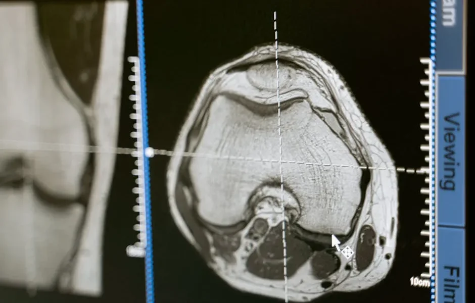
Step 4: Reserve MRI for Brain, Spinal, and Soft Tissue Superiority
MRI (magnetic resonance imaging) sits at the top of the cost pyramid. It costs $2,000-$3,500.
It also provides the most diagnostic sophistication.
It provides 10-15% better soft tissue contrast than CT. This makes invisible problems suddenly crystal clear.
MRI is unmatched for:
- Brain tumors and neurological conditions
- Spinal cord diseases (disc herniations, inflammation)
- Joint ligament and cartilage tears
- Muscle and tendon injuries
- Certain cancers requiring detailed soft tissue evaluation
MRI uses powerful magnets and radio waves. There’s no radiation at all.
But scans take 30-90 minutes. This requires deep general anesthesia throughout.
Any metal in or on your pet can cause problems. This includes some identification microchips. But most modern veterinary microchips are MRI-safe.
The image quality is stunning.
Neurologists can see individual layers of the spinal cord. They can identify tiny brain lesions. They can evaluate soft tissue masses with unparalleled detail.
For a dog suddenly paralyzed from a herniated disc, MRI is often essential. It’s often the only way to pinpoint the exact problem location before emergency surgery.
Not every area has veterinary MRI.
Many practices refer to specialty hospitals or mobile MRI services. Yes, MRI-equipped trucks now serve major metro areas.
This is as of 2024-2025. It’s a development that’s increased access significantly.
Low-field MRI systems (0.3T-0.5T) are emerging for equine imaging. These sometimes allow standing procedures without full anesthesia.
These cost-effective units cost $400K-$600K. High-field systems cost $1.5M or more.
These may eventually become available at more general practices.
Step 5: Match the Modality to the Clinical Question
Here’s where veterinary medicine becomes both art and science.
The “best” imaging depends entirely on what question needs answering.
Let me break down common scenarios:
For Limping and Lameness
Start with X-rays to rule out fractures and obvious joint disease.
If negative but lameness persists, ultrasound can evaluate soft tissue injuries around joints.
For cruciate ligament tears or complex joint problems, CT provides surgical detail.
For muscle or tendon injuries, MRI offers the most information.
For Seizures and Neurological Problems
MRI is the gold standard for brain and spinal cord evaluation.
CT can identify some problems faster and cheaper. These include bleeding and large tumors.
But MRI’s soft tissue detail is unmatched for neurological workup.
For Abdominal Masses or Vomiting
X-rays first for foreign objects and basic survey.
Ultrasound next for organ evaluation and mass characterization.
CT for surgical planning if cancer or complex masses are found.
MRI rarely needed for routine abdominal issues.
For Breathing Difficulties
X-rays are excellent for several things. These include pneumonia, heart disease, and fluid in the chest.
CT provides incredible lung detail. This helps with masses, complex infections, or surgical planning.
Ultrasound can evaluate heart function (echocardiography). But it doesn’t image lungs well.
For Cancer Staging
Often requires multiple modalities.
X-rays or CT for lungs (common metastasis site).
Ultrasound for abdominal spread.
CT or MRI for primary tumor detail. This depends on location.
Your vet’s recommendations should follow a logical diagnostic pathway.
Just like maintaining thorough pet health records, documenting imaging is important. Record which imaging was done and why. This helps track your pet’s medical journey.
Understanding Cost vs. Value in Veterinary Imaging
Let’s talk money. This matters.
An X-ray costs $150-$400.
An MRI costs $2,000-$3,500.
That’s a 10x difference. Is it worth it?
Sometimes absolutely yes.
A dog with sudden paralysis needs MRI immediately. Waiting or trying cheaper options first could mean permanent disability.
Time is literally spinal cord tissue.
But what about a mildly limping dog? One with a small amount of arthritis visible on X-rays?
Jumping straight to MRI would be diagnostic overkill.
The X-ray answered the question and guided treatment.
Here’s what I’ve learned watching thousands of cases:
The “right” imaging done first saves money overall.
Repeat X-rays every few months trying to avoid an inevitable CT scan? You’ll spend the CT cost anyway. It’s just spread out with delayed diagnosis.
Pet insurance typically covers diagnostic imaging when medically necessary. But policies vary.
Some cover only a percentage after deductible. Others have per-incident limits.
Review your policy or ask directly about coverage before expensive imaging.
Many specialty hospitals offer payment plans for advanced imaging.
And mobile MRI services sometimes cost less than hospital-based units.
Don’t be afraid to ask about options if cost is a barrier to necessary diagnostics.
The 2025 Technology Factor: AI and Enhanced Imaging
Veterinary imaging in 2025 isn’t just about the machines.
It’s also about what happens after the image is captured.
AI software like IvyVision and VetCT AI now assist with interpretation. They detect fractures and tumors with over 90% accuracy.
This matters especially at general practices. Board-certified radiologists aren’t on staff at these locations.
AI acts as a safety net. It flags potential abnormalities. The primary vet might review them more carefully. Or they might send them to a specialist.
Contrast agents have also improved.
New veterinary-specific formulations were approved in 2024. They’re safer for pets with kidney disease.
Historically, this was a group that couldn’t safely undergo contrast-enhanced CT or MRI studies.
These aren’t sci-fi futures. They’re happening now. And they’re improving diagnostic accuracy without increasing cost significantly.
Common Tips for Navigating Veterinary Imaging Decisions
I’ve watched countless pet owners navigate these choices. Here’s what actually helps:
- Ask what specific question the imaging will answer: “Will this tell us if surgery is needed?” is clearer than vague “let’s take a look.”
- Understand the diagnostic pathway: Why this test now? What if it’s normal—what’s next? What if it’s abnormal?
- Discuss cost upfront: Good vets appreciate when you’re honest about budget constraints. Sometimes a slightly less ideal but more affordable option still provides useful information.
- Ask about sedation/anesthesia risks: Especially for older pets or those with health issues, anesthesia risk may influence imaging choices. Quick CT might be safer than long MRI for some patients.
- Get copies for your records: Digital images can be transferred between vets. If you’re seeking a second opinion or moving, having your imaging saves repeating expensive tests.
- Consider timing: Emergency imaging availability varies. Most general practices have X-ray, many have ultrasound, few have CT/MRI. Emergency hospitals typically have CT but may not have MRI on-site.
- Ask if a specialist should interpret: Complex cases benefit from board-certified radiologist review, even if imaging is done at your regular vet.
This is similar to evaluating vet nutrition advice.
Don’t be afraid to ask questions about imaging recommendations. Good vets welcome engaged, informed pet owners.
Mistakes to Avoid When Making Imaging Decisions
I’ve seen these costly errors repeatedly:
- Refusing recommended imaging due to cost, then spending more treating blindly: Guessing at treatment without proper diagnosis often costs more long-term than the imaging would have.
- Insisting on advanced imaging when basic tests haven’t been done: Jumping to MRI without X-rays first usually isn’t logical or cost-effective.
- Choosing imaging based solely on price: The cheapest option that doesn’t answer the clinical question wastes money entirely.
- Not asking about anesthesia requirements upfront: Surprises at check-in cause unnecessary stress and sometimes force postponement.
- Failing to follow up on borderline findings: “Recheck in 6 weeks” isn’t optional—monitoring changes over time provides crucial information.
- Assuming mobile/phone consultations can replace imaging: Some diagnoses absolutely require seeing inside your pet. Telemedicine has limits.
- Not considering emergency facility imaging availability: Know before crisis whether your local emergency hospital has CT/MRI or only X-ray and ultrasound.
This is just like understanding responsible medication use.
Informed imaging decisions require balancing multiple factors. These include medical necessity, safety, cost, and urgency.
When to Ask for a Specialist or Second Opinion
Sometimes your general practice vet’s imaging recommendation warrants specialist input.
Consider requesting referral or consultation when:
- Advanced imaging (CT/M
Medical disclaimer: This article is for educational purposes only and does not substitute professional veterinary advice, diagnosis, or treatment. Always consult a licensed veterinarian about your pet's health.